Abstract
Research Article
Comparison of muscle activation of 3 different hip belt squat techniques
Colleen N Gulick* and Dawn T Gulick
Published: 15 September, 2020 | Volume 4 - Issue 2 | Pages: 034-039
The purpose of this study was to differentiate between muscular activity of three different types of belt squats (SquatMax-MD, Pit Shark and Monster Rhino) and the muscle activation of the rectus femoris, vastus medialis oblique, gluteus maximus, and gluteus medius. Fourteen healthy, male athletes, over the age of 18 years, performed 2 sets of 5 repetitions on each of the three belt squat machines with a weight equivalent to each participant’s body weight. Athletes were given at least 2 minutes of rest between each set and condition. Electromyographic data were collected from four muscles: rectus femoris, vastus medialis oblique, gluteus maximus, and gluteus medius muscles. ANOVA revealed the SquatMax-MD belt squat resulted in the highest muscle activation in every muscle, with significantly higher activity in the rectus femoris, vastus medialis oblique, and gluteus medius muscles. The Monster Rhino belt squat produced the second highest muscle activation with the Pit Shark belt squat creating the lowest muscle activation. In totality, the SquatMax-MD produced 38.7% greater muscle activation than the Monster Rhino and 12.2% greater activation than the Pit Shark. The belt squat can be an advantageous exercise because it can effectively load the lower body while de-loading the spine and upper body. The difference in activation between the SquatMax-MD and other belt squats may be due, in part, to the design of the machines. The additional activation produced by the SquatMax-MD belt squat may be useful for individuals seeking hypertrophy, strength, or a reduction in injury risk.
Read Full Article HTML DOI: 10.29328/journal.jnpr.1001035 Cite this Article Read Full Article PDF
Keywords:
Squat; Belt squat; Muscle recruitment; EMG
References
- Paoli A, Marcolin G, Petrone N. The effect of stance width on the electromyographical activity of either superficial thigh muscles during back squat with different bar loads. J Strength Cond Res. 2009; 23: 246-250. PubMed: https://pubmed.ncbi.nlm.nih.gov/19130646/
- Dionisio V, Almeida G, Duarte M, Hirata R. Kinematic, kinetic & EMG patterns during downward squatting. J Electromyogr Kinesiol. 2008; 18: 134-143. PubMed: https://pubmed.ncbi.nlm.nih.gov/17029862/
- Comfort P, Kasim P. Optimizing Squat Technique. Strength and Conditioning J. 2007; 29: 10-13.
- Gulick D, Fagnani J, Gulick C. Comparison of muscle activation of hip belt squat and barbell back squat techniques. Isokinet Exerc Sci. 2015; 23: 101-108.
- Evans T, McLester C, Howard J, McLester J, Calloway J. Camparison of Muscle Activation Between Back Squats and Belt Squats. J Strength Cond Res. 2019; 33: S52-S59. PubMed: https://pubmed.ncbi.nlm.nih.gov/28595237/
- Joseph L, Reilly J, Sweezey K, Waugh R, Carlson L, et al. Activity of Trunk and Lower Extremity Musculature: Comparison Between Parallel Back Squats and Belt Squats. J Human Kinetics. 2020; 72: 223-228. PubMed: https://pubmed.ncbi.nlm.nih.gov/32269663/
- Florimond V. Basics of Surface ELectromyography: Applied to Physical Rehabilitation & Biomechanics. Thought Technology Ltd. 2010; 18-19.
- Gullett J, Tillman M, Gutierrez G, Chow J. A biomechanical comparison of back and front squats in healthy trained individuals. J Strength Cond Res. 2009; 23: 284-292. PubMed: https://pubmed.ncbi.nlm.nih.gov/19002072/
- Basmajian J, Deluca C. Muscle Alive. Baltimore: Williams & Wilkins. 1985.
- Pereira G, Leporace G, Das Virgens Chagas D, Furtado L, Praxedes J, et al. Influence of hip extrenal rotation on hip adductor & rectus femoris myoelectric activity during a dynamic parallel squat. J Strength Cond Res. 2010; 24: 2749-2754. PubMed: https://pubmed.ncbi.nlm.nih.gov/20651607/
- Escamilla R, Fleisig G, Zheng N, Lander J, Barrentine S, et al. Effects of technique variations on knee biomechanics during the squat and leg press. Med Sci Sport Exer. 2001; 33: 1552-1566. PubMed: https://pubmed.ncbi.nlm.nih.gov/11528346/
- Schwanbeck S, Chilibeck P, Binsted G. A comparison of free weight squat to Smith machine squat using electromyography. J Strength Cond Res. 2009; 23: 2588-2591. PubMed: https://pubmed.ncbi.nlm.nih.gov/19855308/
- Wilk K, Escamilla R, Fleisig G, Barrentine S, Andrews J, et al. A comparison of tibiofemoral joint ofrces and electromyographic activity during open and closed kinetic chain exercises. Am J Sports Med. 1996; 24: 518-527. PubMed: https://pubmed.ncbi.nlm.nih.gov/8827313/
- Bolgla L, Malone T, Umberger B, Uhl T. Hip strength and hip knee kinematics during stair descent in females with and without patellofemoral pain syndrome. J Orthopedic Sports Physical Therapy. 2008; 38: 12-18. PubMed: https://pubmed.ncbi.nlm.nih.gov/18349475/
- Fauth MG, Lutsch B, Gray A, Szalkowski C, Wurm B, et al. Hamstrings, quadriceps, & gluteal muscle activation during resistance training exercises, in: ISBS Conference. 2010.
- Myer G, Chu D, Brent J, Hewett T. Trunk & hip control neuromuscular training for prevention of knee injury. Clin Sports Med. 2008; 27: 425-448. PubMed: https://pubmed.ncbi.nlm.nih.gov/18503876/
- Piva S, Goodnite E, Childs J. Strength around the hip and flexibility of soft tissue in individuals with and without patellofemoral pain syndrome. J Orthop Sports Phys Ther. 2005; 35: 793-801. PubMed: https://pubmed.ncbi.nlm.nih.gov/16848100/
- Prins M, Van der Wurff P. Females with patellofemoral pain syndrome have weak hip muscles: A systematic review. Aust J Physiother. 2009; 55: 9-15. PubMed: https://pubmed.ncbi.nlm.nih.gov/19226237/
- Robinson R, Nee R. Analysis of hip strength in females seeking physical therapy treatment for unilateral patellofemoral pain syndrome. J Orthop Sports Phys Ther. 2007; 37: 232-238. PubMed: https://pubmed.ncbi.nlm.nih.gov/17549951/
- Khayambashi K, Ghoddosi N, Straub R. Hip Muscle Strength Predicts Noncontact Anterior Cruciate Ligament Injury in Male and Female Athletes: A Prospective Study. Am J Sports Med. 2015; 44: 355-361. PubMed: https://pubmed.ncbi.nlm.nih.gov/26646514/
- Beardsley C. How important are the hamstrings during squats? Strength & Conditioning Research, 2013.
- Ebben W, Long N, Pawlowski Z, Chimielewski L, Clewien R, et al. Using squat repetition maximum testing to determine hamstring resistance training exercise loads. J Strength Cond Res. 2010; 24: 293-299. PubMed: https://pubmed.ncbi.nlm.nih.gov/20072071/
- Hung Y, Gross M. Effect of Foot Position on Electromyographic Activity of the Vastus Medialis Oblique and Vastus Lateralis During Lower-Extremity Weight-Bearing Activities. J Orthop Sports Phys Ther. 1999; 29: 93-105. PubMed: https://pubmed.ncbi.nlm.nih.gov/10322584/
- Murray N, Cipriani D, O'Rand D, Reed-Jones R. Effects of Foot Position during Squatting on the Quadriceps Femoris: An Electromyographic Study. Int J Exercise Sci. 2013; 6: 114-125. PubMed: https://pubmed.ncbi.nlm.nih.gov/27293497/
Figures:
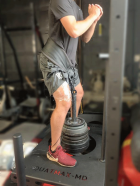
Figure 1
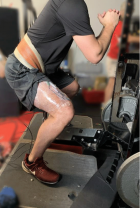
Figure 2
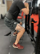
Figure 3
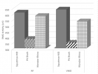
Figure 4
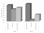
Figure 5
Similar Articles
-
Effects of Fast-Walking on Muscle Activation in Young Adults and Elderly PersonsCamila Fonseca de Oliveira*,Denise Paschoal Soares,Michel Christian Bertani,Leandro José Rodrigues Machado,João Paulo Vila-Boas. Effects of Fast-Walking on Muscle Activation in Young Adults and Elderly Persons. . 2017 doi: 10.29328/journal.jnpr.1001002; 1: 012-019
-
Perceptive and Rehabilitative Muscle Recruitment Facilitation Secondary to the use of a Dynamic and Asymmetric Spine Brace in the treatment of Adolescent Idiopathic Scoliosis (AIS)Maurizio Falso*, Laura Forti,Silvia Iezzi,Gloria Cottali,Eleonora Cattaneo,Marco Zucchini,Franco Zucchini. Perceptive and Rehabilitative Muscle Recruitment Facilitation Secondary to the use of a Dynamic and Asymmetric Spine Brace in the treatment of Adolescent Idiopathic Scoliosis (AIS). . 2018 doi: 10.29328/journal.jnpr.1001021; 2: 054-079
-
Comparison of muscle activation of 3 different hip belt squat techniquesColleen N Gulick*,Dawn T Gulick. Comparison of muscle activation of 3 different hip belt squat techniques. . 2020 doi: 10.29328/journal.jnpr.1001035; 4: 034-039
Recently Viewed
-
Prediction of neonatal and maternal index based on development and population indicators: a global ecological studySedigheh Abdollahpour,Hamid Heidarian Miri,Talat Khadivzadeh*. Prediction of neonatal and maternal index based on development and population indicators: a global ecological study. Clin J Obstet Gynecol. 2021: doi: 10.29328/journal.cjog.1001096; 4: 101-105
-
A Genetic study in assisted reproduction and the risk of congenital anomaliesKaparelioti Chrysoula,Koniari Eleni*,Efthymiou Vasiliki,Loutradis Dimitrios,Chrousos George,Fryssira Eleni. A Genetic study in assisted reproduction and the risk of congenital anomalies. Clin J Obstet Gynecol. 2021: doi: 10.29328/journal.cjog.1001095; 4: 096-100
-
Leiomyosarcoma in pregnancy: Incidental finding during routine caesarean sectionToon Wen Tang*,Phoon Wai Leng Jessie. Leiomyosarcoma in pregnancy: Incidental finding during routine caesarean section. Clin J Obstet Gynecol. 2021: doi: 10.29328/journal.cjog.1001094; 4: 092-095
-
Adult Neurogenesis: A Review of Current Perspectives and Implications for Neuroscience ResearchAlex, Gideon S*,Olanrewaju Oluwaseun Oke,Joy Wilberforce Ekokojde,Tolulope Judah Gbayisomore,Martina C. Anene-Ogbe,Farounbi Glory,Joshua Ayodele Yusuf. Adult Neurogenesis: A Review of Current Perspectives and Implications for Neuroscience Research. J Neurosci Neurol Disord. 2024: doi: 10.29328/journal.jnnd.1001102; 8: 106-114
-
Late discover of a traumatic cardiac injury: Case reportBenlafqih C,Bouhdadi H*,Bakkali A,Rhissassi J,Sayah R,Laaroussi M. Late discover of a traumatic cardiac injury: Case report. J Cardiol Cardiovasc Med. 2019: doi: 10.29328/journal.jccm.1001048; 4: 100-102
Most Viewed
-
Evaluation of Biostimulants Based on Recovered Protein Hydrolysates from Animal By-products as Plant Growth EnhancersH Pérez-Aguilar*, M Lacruz-Asaro, F Arán-Ais. Evaluation of Biostimulants Based on Recovered Protein Hydrolysates from Animal By-products as Plant Growth Enhancers. J Plant Sci Phytopathol. 2023 doi: 10.29328/journal.jpsp.1001104; 7: 042-047
-
Sinonasal Myxoma Extending into the Orbit in a 4-Year Old: A Case PresentationJulian A Purrinos*, Ramzi Younis. Sinonasal Myxoma Extending into the Orbit in a 4-Year Old: A Case Presentation. Arch Case Rep. 2024 doi: 10.29328/journal.acr.1001099; 8: 075-077
-
Feasibility study of magnetic sensing for detecting single-neuron action potentialsDenis Tonini,Kai Wu,Renata Saha,Jian-Ping Wang*. Feasibility study of magnetic sensing for detecting single-neuron action potentials. Ann Biomed Sci Eng. 2022 doi: 10.29328/journal.abse.1001018; 6: 019-029
-
Pediatric Dysgerminoma: Unveiling a Rare Ovarian TumorFaten Limaiem*, Khalil Saffar, Ahmed Halouani. Pediatric Dysgerminoma: Unveiling a Rare Ovarian Tumor. Arch Case Rep. 2024 doi: 10.29328/journal.acr.1001087; 8: 010-013
-
Physical activity can change the physiological and psychological circumstances during COVID-19 pandemic: A narrative reviewKhashayar Maroufi*. Physical activity can change the physiological and psychological circumstances during COVID-19 pandemic: A narrative review. J Sports Med Ther. 2021 doi: 10.29328/journal.jsmt.1001051; 6: 001-007

HSPI: We're glad you're here. Please click "create a new Query" if you are a new visitor to our website and need further information from us.
If you are already a member of our network and need to keep track of any developments regarding a question you have already submitted, click "take me to my Query."


